Introduction
In human gait, the ankle-foot complex plays a crucial role, performing movements that absorb loads imposed on the lower limb and accumulate elastic potential for propulsion [1, 2]. this physiological movement can occur in an excessive way and its main characteristics are initial contact with an excessively pronated hindfoot, a load response and midstance with excessive midfoot depression, a terminal stance and a pre-swing with an excessive supinated forefoot [1, 2].
Various methods are currently employed for detecting functional alterations in the foot, including both dynamic and static assessments. Dynamic analysis relies on sensors that measure foot plantar pressures [3] or three-dimensional motion analysis systems to obtain accurate and reliable measurements [4, 5]. For instance, the Vicon System, recognised as the gold standard for gait assessment and equipped with the Oxford Foot Model Plug-in, is widely employed in the literature for evaluating the movement of foot segments (leg, hindfoot, midfoot, and forefoot) [4–7].
Static analysis is extensively utilised in clinical settings owing to its speed and cost effectiveness. Furthermore, it serves as a screening tool in numerous clinical research studies [2, 8–11]. the gold standard for this evaluation is radiographic classification [2]; however, it poses radiation exposure risks to the patient. Another widely used assessment tool for this purpose is the Foot Posture Index (FPI) [11–14]. the FPI utilises six criteria for evaluation, including palpation and observation of the hindfoot, midfoot, and forefoot, resulting in a classification between supinated, neutral, and pronated feet [11, 12].
Abnormal movement patterns, such as excessive forefoot supination, arch lowering, and excessive hind-foot pronation, can lead to injuries [2], such as medial tibial stress syndrome [1, 13] and foot pain [15]. Excessive pronation may also increase knee internal rotation and valgus [16, 17], potentially leading to anterior knee pain [18]. Moreover, it can also affect the anterior-posterior pelvic tilt [16].
The link between these clinical conditions and excessive foot motion highlights the need for interventions to minimise the risk of injury. In this context, there are several therapeutic options, including insoles [19], therapeutic exercises [20] and therapeutic tape [21]. the most extensively studied tapes are the rigid and elastic ones. Both can be applied with the purpose of correcting excessive foot motion. Rigid tape may reduce pronation, mainly at hindfoot [22, 23]. However, this type of tape tends to restrict the overall foot movement [23].
On the other hand, elastic tape, also known as Kinesio tape, Kinesiology tape, or Neuromuscular tape, may offer less restriction of foot movement, provide mechanical support, and be more comfortable [21, 24]. However, its effects on foot motion remain unclear [14, 25]. to date, no study has investigated the effects of elastic tape on foot motion with a three-dimensional multi-segment foot model in gait assessment. there-fore, the purpose of this study is to examine the influence of elastic tape on the forefoot, midfoot and hind-foot motion of young women.
Previous studies employing rigid or elastic tape have shown some effects in the plantar pressure of the foot, suggesting a degree of control in arch lowering or in the movement of the hindfoot and forefoot [14, 22, 23, 25]. We hypothesise that elastic tape can modify foot movement, similar to what some previous studies have observed in relation to plantar pressure.
Materials and methods
Design
A self-controlled clinical trial was conducted with a blinded evaluator (he did not know which side the tape was applied to during the study).
Subjects
The initial number of participants was 15, however 5 dropped out during the trial. the final sample was 10 female participants, and their descriptive characteristics are shown in Table 1.
Table 1
Sample characteristics
Volunteers were recruited using an electronic poster published on social media. the announcement was addressed to young participants between 18 and 30, with a focus on people who noticed changes in the sole of the foot during gait. the inclusion criteria were: age between 18 and 30 years, body mass index between 18.6 and 24.9 kg/m2, and at least one flat foot assessed with the Foot Posture Index (FPI3 6) [11]. Participants means: experimental side 8 ± 2 and control side 7 ± 2. the FPI utilises six items to determine the foot posture. these items evaluate the positioning of the fore-foot, midfoot, and hindfoot. Each item is scored on a scale from -2 to 2, yielding a total score range of –12 to 12. A value of 6 or greater indicates a flatfoot posture [11]
The exclusion criteria were: surgeries and/or trauma in the lower limb during the past six months, known tape allergy, recent or ongoing excessive midfoot pro-nation treatment, skin disorders at the tape site, use of medications that could impair balance, and having consumed alcoholic beverages within 48 hours prior to the assessment.
Intervention
The tape was applied to the most flattened foot (Experimental Side – ES) and the contralateral foot did not receive any intervention (Control Side – CS). Prior to application of the tape, the skin was cleaned with a 70% alcohol solution.
Elastic tape (Kinesioâ taping tex Gold FP) was applied with the foot and ankle pre-positioned in dorsiflexion, inversion and maximal adduction (functional application). the application consisted of an initial anchor (without stretching), an elastic tension zone (maximum stretching corresponding to 5 Newtons) and a final anchor (without stretching). the initial anchor was fixed between the first and fifth metatarsals on the foot dorsum. the elastic tension zone began on the side of the foot, going obliquely through the navicular tuberosity along the anterior tibialis muscle. the final 5 cm anchor was fixed to the leg’s proximal lateral upper third of the leg. the measurement of tape stretching was done according to the novel method proposed by Matheus et al. (2017) [26].
This application was chosen because it is largely used in clinical practice and, with maximum stretching, the tape used has a rigid stop.
Procedures
All data collection was conducted at the motion analysis laboratory of the State Center for Rehabilitation and Readaptation Dr. Henrique Santillo (CRER) in Goiânia, Goiás, Brazil.
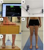
Without and with tape 3D motion data was collected using a Vicon Systemâ, and the Oxford Foot Model (OFM) was used to assess the foot and ankle movement (Figure 1C, 1D, 1E) [4]. Reflective markers with a diameter of 8 mm were affixed using OFM criteria and a laser level for alignment (Figure 1C, 1D, 1E). to capture the kinematic data, four force plates (AMtI® models OR6 and OR7), two VHS cameras, and ten infrared cameras (Pulmix® – model tM 6701AN) were used (Figure 1A, 1B, 1D). the gait analysis data were processed and organised using the Vicon Nexus (Vicon Motion Systems Ltd., Oxford, UK], Vicon Polygon (Vicon Motion Systems Ltd., Oxford, UK) and Microsoft Excel (Microsoft Corporation, Redmond, WA, USA). the trajectories of the markers were sampled at 300Hz, the rate of the force plates was 1200 Hertz, and the filters applied during data processing included: Fill gaps, Butterworth (analogue devices, trajectories and model outputs) and the Woltring filtering routine.
All data collection was done by a single researcher and no marker was removed for tape application. A second researcher, blinded for the study, conducted data processing after the collection phase. the computer model does not display the leg with the applied bandage because it is non-reflective (refer to Figure 1A).
Participants wore close-fitting shorts and walked barefoot along an eight-metre walkway at a self-selected speed. Data collection started after three steps and finished three steps before the end of the walkway. Data from five trials were averaged for each participant and all data were normalised by gait cycle using fifty-one time-normalised samples for each stride.
The following kinematic data was used:
Frontal Plane: supination/pronation of the fore-foot in relation to the hindfoot;
Frontal Plane: supination/pronation of the hind-foot in relation to the leg;
Medial longitudinal arch (MLA) height normalised by the foot length and arch deformation during stance – dimensionless value.
The normalised arch height (A) and arch deformation (B) were calculated as follows: (A) distance between the base of the first metatarsal marker and the plane defined by three forefoot markers (base of the hallux, base of the fifth metatarsal and head of the fifth metatarsal) divided by foot length[4]; (B) arch deformation is the difference between the highest and lowest arch height normalised within the first 10% of the gait cycle[27]. Lower values of arch height indicate a flattened foot and higher values of arch deformation also indicate a flattened foot [4, 27].
Statistical analysis
The sample size was calculated using pilot data from four flatfoot subjects using the G*Power 3.1 software (Heinrich Heine University Düsseldorf, Düsseldorf, Germany). The mean and standard deviation were used for this calculation with and without tape moments, using the following parameters: expected effect size = 1.03, error probability (α ) = 0.05 and power (1 –β ) = 0.95. Thus, the estimated sample size was 15 participants.
Shapiro–Wilk was used to test for a normal distribution. The intragroup comparisons were conducted using the paired Student’s t-test and the Wilcoxon test for non-parametric data. The intergroup comparisons were conducted using the independent t-test and the Mann–Whitney test for non-parametric data. The significance level considered was 0.05. The effect size was calculated as follows: dz = t/√n (dz – effect size, t – the observed value of t test, n – sample size) for parametric data (0.2 – small, 0.5 – medium, 0.8 – large); for non-parametric data r = Z/ √2xn (r – effect size, Z – observed Wilcoxon test, n – sample size) (0.1 – small, 0.3 – medium, 0.5 – large). Statistical analysis was performed using IBM SPSS Statistics version 23 (IBM Corp., Armonk, NY, USA).
Ethical approval
The research related to human use has complied with all the relevant national regulations and institutional policies, has followed the tenets of the Declaration of Helsinki, and has been approved by the Ethics Committee of the Pontifícia Universidade Católica de Goiás – PUC/Goiás (approval No.: 1.450.171, CAAE: 53240515.2.0000.0037). Additionally, this study has been registered with the clinical trial registration code: U1111-1197-4728 (http://www.ensaiosclinicos.gov.br/).
Informed consent
Informed consent has been obtained from all individuals included in this study. All participants provided written informed consent prior to data collection, ensuring adherence to ethical standards.
Results
The characteristics of the sample and spatial-temporal gait data can be found in Tables 1 and 2. the cadence, gait speed, double-support, and single-support durations were not affected by the application of taping. Similarly, the step length and step width for both the experimental and control sides remained unchanged.
Table 2
Spatial-temporal gait data
Intragroup and intergroup comparisons were made for measurements of the forefoot, MLA height and hindfoot (Tables 3 and 4).
Table 3
Forefoot motion, arch height, arch deformation and hindfoot motion without and with elastic tape.
Table 4
Forefoot motion, arch height, arch deformation and hindfoot motion without and with elastic tape.
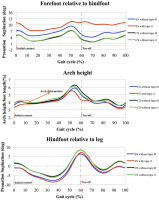
The forefoot motion showed statistically significant differences between the without and with tape conditions during the initial contact (mean difference: ES = 3.82, CS = 0.18), toe-off (mean difference: ES = 5.81, CS = –0.3) and maximum pronation (mean difference: ES = 2.41, CS = 0.01). these differences were significant only in the ES and exhibited a large effect size (greater than 0.8). During the intergroup comparison, the baseline revealed no significant differences, but post-intervention, significant differences were confirmed. Initial contact (mean difference: without tape =2.26, with tape = 5.91), toe-off (mean difference: without tape = 2.14, with tape = 8.24) and maximum pro-nation (mean difference: without tape = 2.54, with tape = 4.94). these findings show an increase in fore-foot supination during the stance phase of gait.
The MLA height showed statistically significant differences between the without and with tape conditions during toe-off (mean difference: ES = –0.73, CS = –0.4) and arch deformation (mean difference: ES = –0.16, CS = –0.25). However, this difference was not significant at initial contact (mean difference: ES = –0.52, CS = –0.83). these differences were significant only on the ES and exhibited a large effect size (greater than 0.8 for parametric and greater than 0.5 for non-parametric). During the intergroup comparison, no significant differences were observed either at the baseline or post-intervention moments. Initial contact (mean difference: without tape = –0.52, with tape = –0.83), toe-off (mean difference: without tape = –0.51, with tape = –0.84) and arch deformation (mean difference: without tape = –0.17, with tape = –0.08).
The hindfoot motion did not exhibit statistically significant differences between the without and with tape conditions during the initial contact (mean difference: ES = 0, CS = 0.36), toe-off (mean difference: ES = 0.95, CS = 0.02) and maximum pronation (mean difference: ES = 0.76, CS = 0.76). During the intergroup comparison, no significant differences were observed either at the baseline or post-intervention moments at initial contact (mean difference: without tape = –2.54, with tape = –2.95), toe-off (mean difference: without tape = –5.09, with tape = –1.34) or maximum pronation (mean difference: without tape = 5.36, with tape = 2.49).
Figure 2 shows the graphs of the ES and CS without and with tape for the forefoot, MLA height and hind-foot. It also shows how the arch deformation was verified.
Discussion
The aim of the study was to investigate the influence of elastic tape on flatfoot motion using a 3D multi-segment kinematic assessment. to accomplish this, fore-foot supination/pronation [22], arch height [2], arch deformation [27] and hindfoot supination/pronation [28] were chosen as important measures. to assess these measures, we used the outputs of the OFM. Several studies have evaluated the reproducibility of this biomechanical model, mainly assessing healthy children [4, 29]. Moderate to good intraclass correlation coefficients (ICC) for inter-rater and intra-rater reliability were found for hindfoot and forefoot movements in healthy adults [7]. No study was found assessing the ICC for OFM arch height. to improve the reproducibility, no reflective marker was removed for tape application and all evaluations were done by a single investigator.
This investigation revealed three primary findings: an increase in forefoot supination during initial contact, toe-off, and maximum pronation in the stance phase; a decrease in arch height during toe-off; and a reduction in arch deformation during the loading response. No changes were observed in the hindfoot movement.
To the best of the authors’ knowledge, no study has been performed to date that assesses the movement of the ankle-foot complex in both the sagittal and frontal planes using a multi-segment foot model and comparing without and with the application of elastic tape. Due to this innovative feature, our results will be discussed against similar studies that used different investigation methods.
Other authors have found similar results showing a reduction in the medial forefoot and midfoot loading rate with no hindfoot effect when assessing subjects with medial tibial stress syndrome after elastic tape application with 75% tension [30].
In contrast, two studies assessing plantar pressure found no difference in foot loading after the application elastic tape [25, 31]. the first [25] did not differentiate the medial and lateral surfaces of each segment, a necessary step to compare with the frontal plane movement evaluated in our study. Also, their tensioned group showed more foot posture correction than their tension-free group, thus agreeing with our findings. the second study [31] employed a different elastic tape than ours, which did not have a maximum tension limit (hyperelastic tape).
A double-blind, randomised clinical trial found no difference in foot posture between elastic tape application in the hindfoot with 100% tension and no tension [14]. two points of discussion are highlighted in this article: first, the application of elastic tape only in the hindfoot segment, and second, the absence of a comparison between the moments before and after the interventions.
Another investigation, that used elastic tape applied with 115–120% tension on the hindfoot and midfoot did not observe a decrease in the medial pressure of the forefoot. However, it did report a decrease in the me-dial pressure of the midfoot and hindfoot[10]. thus, application of elastic tape on the hindfoot and midfoot affects these specific sites[10,25]. Similarly, when the tape is applied to the anterior foot, it leads to a greater midfoot and forefoot effect [30].
One possible explanation for the increased fore-foot supination and the reduction of arch deformation could be related to the lower limb kinetics. Previous research has found an increase in the timing of the peak mediolateral ground reaction during the landing of a jump, following the application of the elastic tape to the leg and hip [32]. this indicates that there was an improvement in the load absorption capacity, indicating that the tape had an influence on the movement in the frontal plane.
It should be emphasised that forefoot supination and arch height are clinically relevant. this is supported by research that also used OFM and verified that these measurements differed between symptomatic flatfoot and typically developing feet [1]. Arch deformation is important in a clinical context as well, since it is a comparable variable to determine the treatment effects of rigid tape in flatfeet [27].
Another result found in this study is the decrease in the arch height and arch deformation on the toe-off and loading response, respectively. Studies evaluating arch behaviour after the application of rigid tape found decreased arch deformation [27] but not decreased arch height [27, 33].
A possible explanation for the decrease in arch height is related to the change of movement that occurred in the foot. Our findings revealed increased forefoot supination after tape application without hind-foot effects, which is a torsional mechanism according to [1, 5]. therefore, we believe that the lowered foot-arch in our results is a consequence of increased forefoot supination since the OFM uses a forefoot reference to measure the foot-arch. Our findings also revealed a decrease in arch deformation during the loading response. Nevertheless, in intergroup comparisons, the experimental side did not show improvement when compared to the control side.
Supporting this perspective, another study utilising a different type of tape (hyperplastic – lacking a rigid stop at the end of stretching) demonstrated similar results: improvement of forefoot supination movement and reduction in arch height. However, unlike the observations made with this elastic tape, there was no intragroup or intergroup decrease in arch deformation [34].
Therefore, elastic tape applied with maximum tension appears to exert an external support effect, in terms of increasing forefoot supination. It is speculated that these effects could be clinically exploited for short-term objectives to provide external support to the foot, promoting increased supination. It is important to remember that the tape is an auxiliary resource and will work best with therapeutic exercise [9, 35, 36]. Caution is necessary, as this study did not investigate the effects after the removal of the tape.
This study has several limitations that should be acknowledged, starting with its small sample size. there also was no long-term evaluation. to achieve an effect closer to clinical reality, the tape was applied on the forefoot and midfoot, which is different from other investigations. It was not possible to assess midfoot motion directly. the height of the normalised MLA lacks reproducibility. Skin movement after tape application might also have biased the results. the screening method employed was the FPI, not radiographic measures. Additionally, no tests were conducted to determine if the subjects’ feet were mobile or rigid. Comparing our findings with previous studies is challenging, particularly due to differences in assessment tools used and the tape tension applied. In most papers, tape tension seems to be applied subjectively, with the tension percentage measured manually.
In a broader field, there is evidence in the literature that other techniques, such as insoles [19] and rigid tapes [22, 23], are also capable of reducing flatfoot characteristics. thus, the choice of therapeutic intervention in clinical practice should consider the patient’s personal preference and practice, type of activity and characteristics [22]. Future investigations should focus on the long-term effects of tape application and lower limb kinetics and should compare elastic tape with rigid tape and insoles.
Conclusions
The application of elastic tape altered the frontal forefoot motion in female participants in the short-term, as it improved the forefoot supination during initial contact, toe-off and maximum pronation. However, the tape did not influence the arch height or frontal hindfoot motion. Future studies must focus on the long-term effects and compare the application with other therapeutic interventions.


